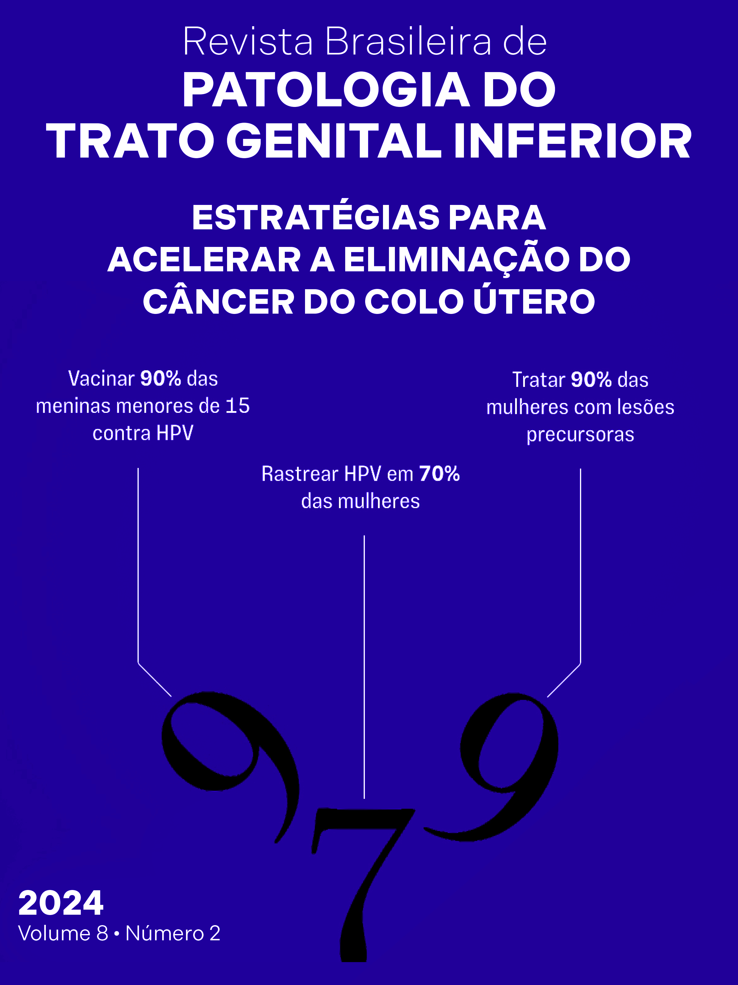
Lesões equívocas em patologia cervical: como intervir?
DOI:
https://doi.org/10.5327/2237-4574-2024820009Palavras-chave:
câncer, colo uterino, diagnósticoResumo
Lesões equívocas são aquelas com comportamento biológico incerto, cujas atipias dificultam a interpretação e impactam
diretamente na abordagem terapêutica, podendo causar atraso no tratamento ou sobretratamento. Um terço dessas lesões
refere-se às atipias anteriormente classificadas como neoplasia intraepitelial grau 2 (NIC2). O epitélio glandular uterino
também apresenta um grande número de lesões equívocas, com atipias que nem sempre indicam neoplasia. Nesses casos, exames
complementares, como a imunoexpressão de p16, são fundamentais para um diagnóstico mais preciso e manejo adequado.
O p16 ajuda a identificar lesões com potencial maligno, sendo especialmente útil na cito e histopatologia. Contudo, sua
expressão anormal não indica prognóstico definitivo, pois a evolução das lesões depende de fatores imunológicos, hormonais
e genéticos. A prática da medicina personalizada, levando em conta o histórico da paciente e seu perfil de risco, é essencial
para decisões mais assertivas e eficazes no tratamento.
Referências
Pruski D, Przybylski M, Millert-Kalinska S, Zmaczynski A, Jach R.
Histopathological discrepancies between colposcopy-directed biopsy
and LEEP-conization observed during SARS-CoV-2 pandemic. Ginekol
Pol. 2023;94(1):12-8. https://doi.org/10.5603/GP.a2022.0081
Maniar KP, Sanchez B, Paintal A, Gursel DB, Nayar R. Role of the
biomarker p16 in downgrading -IN 2 diagnoses and predicting highergrade lesions. Am J Surg Pathol. 2015;39(12):1708-18. https://doi.
org/10.1097/PAS.0000000000000494
Tao AM, Zuna R, Darragh TM, Grabe N, Lahrmann B, Clarke MA, et al.
Interobserver reproducibility of cervical histology interpretation
with and without p16 immunihistoquemistry. Am J Clin Pathol.
;161(2):202-9. https://doi.org/10.1093/ajcp/aqae029
Mody DR, Thrall MJ, Krishnamurthy S. Diagnostic pathology:
cytopathology. 2nd ed. Philadelphia: Elservier; 2018.
Nayar R, Wilbur DC. The pap test and Bethesda 2014. Cancer
Cytopathol. 2015;123(5):271-81. https://doi.org/10.1002/cncy.21521
Staebler A, Sherman ME, Zaino RJ, Ronnet BM. Hormone receptor
immunohistochemistry and human papillomavirus situ hybridization
are usefulfor distinguishing endocervical and endometrial
adenocarcinomas. Am J Surg Pathol. 2002;25(8):998-1006. https://
doi.org/10.1097/00000478-200208000-00004
Waxman AG, Chelmow D, Darragh TM, Lawson H, Moscicki AB.
Revised terminology for cervical histopathology and its implications
for management of high-grade squamous intraepithelial lesions
of the cervix. Obstet Gynecol. 2012;120(6):1465-71. https://doi.
org/10.1097/aog.0b013e31827001d5
Höhn AK, Brambs CE, Hiller GGR, May D, Schmoeckel E, Horn LC. 2020
WHO Classification of Female Genital Tumors. Geburtshilfe Frauenheilkd.
;81(10):1145-53. https://doi.org/10.1055/a-1545-4279
Liao GD, Kang LN, Chen W, Zhang X, Liu XY, Zhao FH, et al. p16
immunohistochemistry interpretation by nonpathologists as an
accurate method for diagnosing cervical precancer and cancer. J
Low Genit Tract Dis. 2015;19(3):207-11. https://doi.org/10.1097/
LGT.0000000000000080
Perkins RB, Smith DL, Jeronimo J, Campos NG, Gage JC, Hansen N,
et al. Use of risk-based cervical screening programs in resourcelimited settings. Cancer Epidemiol. 2023;84:102369. https://doi.
org/10.1016/j.canep.2023.102369
Perkins RB, Wentzensen N, Guido RS, Schiffman M. Cervical cancer
screening: a review. JAMA. 2023;330(6):547-58. https://doi.
org/10.1001/jama.2023.13174

